rsabhk.co.id, 18 September 2024, 12:05 WIB.
Kenapa Penting Melakukan USG ?
USG or ultrasonography is a medical procedure in the form of scanning human organs using high-frequency sound waves. The USG device will then emit high-frequency sound waves and will record the reflected sound waves so that it can display an image of the shape and size of the organ on the monitor screen.
USG can be used to detect various diseases as a supporting tool in surgical processes and also to check pregnancy in women.
Ultrasound examination during pregnancy is useful to support proper and accurate clinical examination of a pregnancy. This examination can obtain some important information such as the location of the pregnancy, identify the number of fetuses being carried, and assist in making prenatal diagnosis decisions in cases of congenital abnormalities in the fetus.
dr. Novan Satya Pamungkas, Sp.O.G, Subsp.K.Fm as an obstetrician and gynecologist subspecializing in maternal and fetal health at RSAB Harapan Kita revealed that USG has become a vital tool for obstetricians, like a modern stethoscope that allows them to monitor the health of the mother and fetus in more detail and accurately. Ease of use and wide availability, even in remote areas, make USG a very helpful tool in providing optimal pregnancy services.
"Today's USG is like a stethoscope for obstetricians. Because it is relatively easy to use and the equipment is widely available, almost all corners now have USG equipment. So, we are greatly helped by the use of this USG to provide services to pregnant women so that their pregnancies can go well and the babies born can be free from complications," said Dr. Novan.
Ultrasound in Early Pregnancy
An ultrasound examination during early pregnancy, after being declared positive through a testpack, has several important roles. First, an ultrasound helps determine the certainty of pregnancy. This is important to ensure that pregnancy is really occurring and not another condition that resembles pregnancy symptoms.
Second, an early ultrasound helps determine the location of the pregnancy. This is important to detect the possibility of a pregnancy outside the uterus (ectopic pregnancy), which requires immediate medical attention. An ectopic pregnancy can be dangerous to the health of the mother and baby if not treated promptly.
Third, early ultrasound helps determine the gestational age more precisely. This is important for calculating the estimated delivery date and monitoring subsequent fetal development. By knowing the accurate gestational age, doctors can provide appropriate advice and recommendations regarding pregnancy examinations and care.
Then by knowing the gestational age from the beginning, it can detect more precise interpretation of labor. USG in the first trimester, the accuracy of predicting gestational age is only off by about 2 to 3 days. However, the greater the gestational age, the greater the error in predicting gestational age. For second trimester USG, pregnancy prediction can be off by 1 to 2 weeks. It can even be off by 3 to 4 weeks if USG is performed in the third trimester.
"So the importance of an ultrasound during early pregnancy from a positive test pack is to determine the certainty of the pregnancy. And then determine the location of the pregnancy, of course we don't want the pregnancy to be outside the uterus. And then from the early ultrasound, we can determine the gestational age more precisely," said Dr. Novan.
"It turns out that the baby has a problem, its growth is slow, even though the gestational age is 20 weeks, but because the growth is slow we predict 16 weeks. Of course this is not right, it will be confusing to determine the gestational age. That's why if there is a patient for control, we always use the first trimester USG," explained Dr. Novan.
In addition, the first trimester of pregnancy, especially the gestational age of 11 to 13 weeks, is a narrow window that offers a golden opportunity to detect possible genetic abnormalities in the fetus. This crucial period is often missed by patients. In this pregnancy period, early detection of genetic abnormalities can be carried out which can have a significant impact on the health and quality of life of the baby.
"Well, what's more important, also in the first trimester at 11 weeks to 13 weeks of pregnancy, we can assess the possibility of genetic abnormalities in the fetus. That window is very, very narrow. Sometimes patients often miss it, even though it is very important at that gestational age. Genetic abnormality syndromes, such as trisomy 21, trisomy 18 and trisomy 13 can be predicted by the first trimester ultrasound examination," explained Dr. Novan.
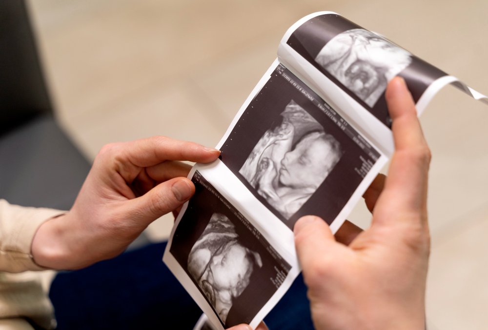
USG 2D, 3D dan 4D
USG 2DUSG 2D, a favorite choice among the three types of pregnancy examinations, is generally performed in the first three months of pregnancy to assess fetal health and detect congenital abnormalities.
Despite displaying monochrome visualization, 2D ultrasound is able to provide detailed images of the developing fetus' internal organs with precision. Through this examination, doctors can measure the length and weight of the fetus, the volume of amniotic fluid, and identify potential fetal abnormalities. The advantages of 2D ultrasound compared to other types of ultrasound are its relatively low cost and wider availability.
USG 3D dan 4D is a development of 2D USG which aims to provide more detailed visualization of pregnancy. This technology allows doctors and mothers to see the baby's face more clearly, even to the structure of internal organs.
USG 3D produces better static (non-moving) three-dimensional images, allowing observation of fetal facial details such as eyes, nose, and lips. This capability helps doctors diagnose fetal organ abnormalities in the womb, such as cleft lip, heart defects, and other congenital abnormalities.
USG 4D has more advanced technology. This ultrasound is able to display three-dimensional images that move in real-time. This scan allows visualization of the fetus from various angles with a higher level of detail. Similar to 3D ultrasound, 4D ultrasound can also diagnose congenital abnormalities.
Many prospective parents wonder whether it is always necessary to use 3D or 4D ultrasound to get a clearer picture of their fetus. Dr. Novan believes that 3D and 4D ultrasound are not always necessary. 2D ultrasound, with good quality, is enough to accurately assess the condition of the fetus.
Furthermore, Dr. Novan explained that 3D and 4D USG do offer more realistic and detailed visualization, but their role is more inclined towards the entertainment aspect, namely to see the baby's face before birth. 2D USG is sufficient for health checks and diagnostic accuracy.
Therefore, there is no need to be fixated on 3D or 4D ultrasound. Consult with your obstetrician to determine the right type of ultrasound according to your needs and pregnancy conditions.
"Does it always have to be with 3D and 4D ultrasound to get better ultrasound accuracy? It's not like that, so with a good quality 2D ultrasound we can also assess the condition of the fetus. It doesn't have to be with 3D or 4D. Because 3D and 4D ultrasound are more for entertainment, to see the baby's face to both parents," said Dr. Novan.
However, Dr. Novan also explained that although 2D ultrasound is generally sufficient for fetal health checks, in some cases, 3D or 4D ultrasound can provide additional benefits. For example, if the obstetrician suspects an abnormality such as a cleft lip. 2D images may be difficult for parents to understand, so more realistic 3D or 4D visualizations can help them better understand the baby's condition.
USG Fetomaternal
The term “fetomaternal ultrasound” is often used by patients, but the correct term is actually “detailed scan ultrasound”. This examination aims to assess the anatomy of the fetus in more detail and comprehensively to detect abnormalities in its organs.
A detailed ultrasound scan is usually performed by a maternal-fetal consultant, an obstetrician who has special expertise in handling high-risk pregnancies and fetal abnormalities.
"Actually, there is no term for fetmaternal USG, what exists is a detailed USG scan. To assess the anatomy of the fetus in more detail to see whether its organs are normal or there are abnormalities. Well, this is called a fetomaternal USG because usually the one who does it is a fetomaternal consultant doctor," said Dr. Novan.
Furthermore, Dr. Novan explained that a detailed USG scan examination is ideally only done once. After the important moment of the detailed USG scan, the focus shifts to monitoring the growth and development of the fetus through a regular USG examination.
"Yes, so a detailed USG scan is actually only enough once. So the 20 to 24 weeks for organ screening is only once. Then after that, just monitoring the growth and development of the fetus is only done with a regular USG examination, no need for a fetomaternal consultant," explained Dr. Novan.
Although a detailed USG scan at 20-24 weeks of pregnancy is ideally performed once, it does not mean that further monitoring is ignored. In the interview, it was stated that the obstetrician will continue to monitor the growth of the fetus, and if there are indications of disorders, a referral back to the fetomaternal consultant will be made. This is because pregnancy is dynamic, where the condition of the fetus can change over time.
"Except if later under monitoring by the obgyn doctor. Why does this growth seem to be disturbed? Pregnancy is dynamic, it could be that at 20 weeks it looks normal, but at 26 weeks there is hydrocephalus, there is fluid in the fetus' head, that's possible. Well, usually later if there is a problem, it will be referred back to the fetomaternal consultant," said Dr. Novan.
Basic Ultrasound By Midwife
The Indonesian Obstetrics and Gynecology Association (POGI) understands the important role of midwives in improving maternal and infant health, including in pregnancy check-ups. Therefore, POGI provides special training for midwives to perform basic ultrasounds.
Midwives who have attended the training and obtained a competency certificate are allowed to perform basic USG. This is in line with the Ministry of Health's goal to reduce the Maternal Mortality Rate (MMR) and Infant Mortality Rate (IMR) in Indonesia.
"So from the POGI (Indonesian Obstetrics and Gynecology Association) organization itself, there is indeed training for midwives. As long as the midwives have undergone the training and have been given a competency certificate to perform basic USG, then please go ahead. It's okay, because the goal is to reduce the Maternal Mortality Rate and Infant Mortality Rate," said Dr. Novan.
Healthy Conclusion
For every couple who wants to have children, getting quality offspring is certainly a dream. In this modern era, the main focus of parents has shifted from quantity to quality. Having healthy and quality children is the main priority.
Preparing for a quality pregnancy begins long before pregnancy. Maintaining maternal health with a nutritious diet, controlling weight, and preventing sexually transmitted diseases are important first steps.
During pregnancy, quality pregnancy check-ups are key. Ultrasound examination, as one of the vital aids, has an important role.
For pregnant women who live in big cities, utilizing the services of a fetomaternal doctor for USG examinations, especially screening tests and second and third trimester screening, is highly recommended. Fetomaternal doctors have special expertise in diagnosing and treating complex pregnancy problems, thus providing peace and certainty for pregnant women and their babies.
Speaker: Dr. Novan Satya Pamungkas, Sp.O.G, Subsp.K.Fm